
Serpin family protease inhibitors (SERPINs) in protozoans are key regulators of multiple biological functions, including the entry of parasites into host cells. Despite their importance, the molecular mechanisms by which SERPINs facilitate host invasion remain largely unresolved. Here, we demonstrate that Eimeria tenella SERPIN1 (EtSERPIN1), present on the sporozoite surface, plays a functional role in cell adhesion and invasion. To identify the host membrane component mediating EtSERPIN1-driven invasion, we applied GST pull-down and yeast two-hybrid approaches, revealing a specific interaction with annexin A2 (ANXA2). Recombinant GgANXA2 bound to sporozoites effectively, and pre-treatment of host cells with either anti-GgANXA2 antibody or recombinant GgANXA2 protein significantly inhibited EtSERPIN1 attachment. Furthermore, recombinant GgANXA2 reduced sporozoite infection in DF-1 cells and in chickens. These findings indicate that the EtSERPIN1–GgANXA2 interaction is critical for E. tenella sporozoite adhesion and invasion. Finally, functional assays with recombinant proteins showed that GgANXA2 significantly decreased sporozoite infectivity, as shown by lower parasite loads in the chicken cecum. Immunization with recombinant EtSERPIN1 provided strong protective effects, including improved body weight gain, reduced cecal lesions and oocyst shedding, and elevated mucosal antibodies. This study highlights EtSERPIN1 as a promising candidate for therapeutic targeting via the GgANXA2 pathway.
Introduction
Avian coccidiosis, caused by infection with one or multiple Eimeria species, leads to severe economic losses globally [1]. Control strategies rely largely on anticoccidial drugs and live vaccines, but drug use is increasingly limited due to resistance, residues, regulations, and delivery challenges in feed [2, 3]. The use of live Eimeria vaccines is also constrained by high production costs and potential pathogenicity [4]. Understanding parasite-host interactions and invasion mechanisms is critical for developing novel intervention strategies.
Host cell invasion by Eimeria involves four steps: attachment, apical reorientation, moving junction formation, and parasitophorous vacuole formation. Proteins secreted from the apical organelles of the parasite mediate these events [5]. Microneme proteins (MICs) have been identified as essential for host cell attachment, using domains such as apple, MIC adhesive repeat regions (MARR), integrin-like A, lectin, and EGF-like domains [6–11]. However, knockout experiments indicate that MICs alone do not fully prevent sporozoite invasion [12, 13], suggesting additional molecules participate in adhesion. One such group is SERPIN protease inhibitors, implicated in facilitating parasite-host attachment and entry [14, 15].
SERPINs are a highly conserved protein family found across viruses, protozoa, and mammals [16]. They act as serine protease inhibitors and regulate processes such as coagulation, fibrinolysis, angiogenesis, apoptosis, development, and immune responses [17]. The first protozoan SERPIN identified was Toxoplasma gondii SERPIN1 (TgPI) [15, 18, 19], followed by discoveries in Neospora caninum, Entamoeba histolytica, and Eimeria spp. [14, 15, 18, 20]. In Eimeria, SERPINs have been reported in E. tenella and E. acervulina [14, 15], and are linked to sporozoite invasion, although their precise role remains unclear. This study aimed to identify host membrane proteins interacting with EtSERPIN1 via pull-down and mass spectrometry, to uncover the molecular mechanism of EtSERPIN1-mediated invasion.
Materials and Methods
Plasmids, yeast, parasites, cells, and animals
We used pCTCON2 plasmid and Saccharomyces cerevisiae EYB100 stored in our lab for yeast surface display and adhesion assays [11]. Wild-type E. tenella Shandong strain-01 (SD-01) was isolated and maintained in-house [21]. DF-1 chicken embryo fibroblast cells were cultured in DMEM (Gibco, USA) with 10% FBS (Biological Industries, Israel) and 1% penicillin-streptomycin, at 37 °C in 5% CO₂. Mycoplasma contamination was excluded using a detection kit (Cat# CA1080, Solarbio, Beijing, China).
BALB/c mice (Jinan Pengyue Experimental Animal Breeding Co., Ltd) were used to generate polyclonal antibodies (pAb) as described [21]. Coccidia-free, 1-day-old Hy-Line layer chicks were purchased from Dongyue Breeder Company (Tai’an, China) and housed under sterile conditions with antibiotic-free feed and water.
Subcellular localization of EtSERPIN1
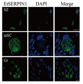
Indirect immunofluorescence assay (IFA) was conducted to determine EtSERPIN1 localization in E. tenella. Sporozoites, second-generation schizonts, and gametocytes were fixed, permeabilized, and incubated sequentially with anti-EtSERPIN1 pAb and FITC-conjugated goat anti-mouse IgG (Solarbio). Nuclei were stained with DAPI (Solarbio). Imaging was performed using a Nikon-ECLIPSE fluorescence microscope (Japan).
Sporozoite secretion assay
To begin, sporulated oocysts were subjected to sterilization by immersing them in a 20% sodium hypochlorite solution for 10 minutes at room temperature, effectively eliminating potential contaminants. Following this treatment, the oocysts were mechanically broken open by vigorous grinding with glass beads, allowing the release of sporocysts contained within. For excystation, the sporocysts were incubated in a sterile phosphate-buffered saline (PBS) solution supplemented with 0.25% trypsin (Solarbio) and 0.25% sodium taurocholate at 41 °C, with gentle agitation, for 40–60 minutes. The liberated sporozoites were collected by filtration using a G3 funnel, ensuring removal of residual sporocyst debris. To trigger secretion of sporozoite proteins, the collected sporozoites were then incubated with 400 nM calcium ionophore A23187 (Aladdin, Shanghai, China) at 37 °C for 20 minutes. After incubation, the supernatant containing the secreted proteins was harvested by centrifugation at 1000 rpm and subsequently stored for downstream protein analyses. This procedure ensured that the proteins released during sporozoite activation could be reliably assessed in later experiments.
Glutathione S‑Transferase (GST) Pull-Down assay
Extraction of Chicken Cecal Membrane Proteins: Cecal epithelial tissue was carefully scraped from healthy chickens and immediately stored at -80 °C to preserve protein integrity. Membrane proteins were then isolated using the Membrane Protein Extraction Kit (Solarbio) in accordance with the manufacturer’s instructions, ensuring enrichment of the membrane-associated fraction for interaction studies.
Pull-Down Procedure: Recombinant GST-tagged EtSERPIN1 (rGST-EtSERPIN1) protein was expressed in E. coli BL21 cells, purified, and incubated with glutathione-conjugated resins (Solarbio) at 4 °C for 5 hours to immobilize the bait protein. A GST-only protein was similarly incubated with resins as a negative control. Following three consecutive washes to remove non-specifically bound proteins, the resins were incubated with the previously extracted chicken cecal membrane proteins at 4 °C for 2 hours, allowing potential binding partners to associate with EtSERPIN1. The complexes were then eluted using 10 mM reduced glutathione (pH 8.0). Finally, all samples were separated by 12% SDS-PAGE, enabling visualization of protein complexes prior to identification.
Mass spectrometry analysis
Protein bands obtained from the pull-down assay were subjected to silver staining using the Fast Stain Silver Kit (Beyotime, Shanghai, China) following manufacturer instructions. Bands unique to the rGST-EtSERPIN1 lane were carefully excised and subjected to tryptic digestion. Peptides were analyzed using a QSTAR XL mass spectrometer (Applied Biosystems, CA, USA), following a standardized protocol. Mass spectrometry was conducted by BPI Genomics (Shenzhen, China), ensuring high-confidence identification of proteins interacting with EtSERPIN1.
Yeast two-hybrid (Y2H) validation
The coding sequence of EtSERPIN1 was inserted into pGBKT7 plasmid to create the bait construct. For prey constructs, coding sequences of chicken annexin genes GgANXA2, GgANXA3, GgANXA5, and GgANXA13 were individually cloned into pGADT7 plasmid. Bait and prey constructs were co-transformed into Y2H Gold strain using the Y2HGold-GAL4 interaction kit (Coolaber, Beijing, China). Transformants were plated on selective media (DDO/X/A and QDO/X/A) and incubated at 30 °C for 3–5 days. Growth on selective plates indicated positive protein-protein interactions. The well-characterized pGBKT7-p53/pGADT7-T pair was used as a positive control to validate the assay.
Western blot analysis
Western blotting was performed to confirm protein interactions identified in the GST pull-down assay. Samples, including both isolated proteins and pull-down complexes, were separated by 12% SDS-PAGE and transferred onto PVDF membranes (Millipore). Membranes were probed with specific antibodies, and protein bands were visualized using NcmECL Ultra substrate (New Cell and Molecular Biotech, Suzhou, China) following the manufacturer’s instructions. This approach allowed confirmation of the presence of EtSERPIN1-interacting proteins and verification of the pull-down results.
Yeast surface display adhesion assay
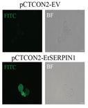
The adhesion capacity of EtSERPIN1 was examined using a yeast surface display system, as described by Wang et al. [22]. In brief, DF-1 cells were incubated with 100 EtSERPIN1-displaying yeast cells for 2 hours at 30 °C. Non-adherent yeast cells were removed by washing three times with PBS, and the remaining yeast were plated on SDCAA medium (0.67% yeast extract, 2% glucose, 0.5% casein acid hydrolysate, 0.15% agar powder, 10% ampicillin, 0.8% agar). After 24 hours, colonies were counted to determine the adhesion rate. Yeast transfected with empty pCTCON2 plasmid served as a control.
Adhesion Inhibition Assay: Two strategies were employed: (1) Yeast displaying EtSERPIN1 were pre-incubated with varying concentrations of recombinant His-tagged GgANXA2 (rHis-GgANXA2) for 1 hour prior to DF-1 exposure; (2) DF-1 cells were pre-treated with anti-ANXA2 polyclonal antibodies, followed by incubation with EtSERPIN1-displaying yeast. Negative controls included PBS or healthy IgG-treated cells. All experiments were conducted in triplicate to ensure reproducibility.
EtSERPIN1 binding to chicken cecum
To examine whether EtSERPIN1 binds to the chicken cecum, an immunohistochemical assay was performed [23]. Cecal tissue was first rinsed thoroughly with sterile PBS, then fixed in 4% paraformaldehyde and embedded in paraffin. Sections were subjected to heat-induced antigen retrieval by boiling in sodium citrate buffer (Servicebio, Wuhan, China) for 10 minutes, followed by blocking with TBST (tris-buffered saline with 0.05% Tween-20) containing 5% BSA to prevent non-specific antibody binding. Tissue slices were then incubated with either recombinant His-tagged EtSERPIN1 (rHis-EtSERPIN1) or PBS as a control at 37 °C for 1 hour. Following this, sections were exposed overnight at 4 °C to anti-EtSERPIN1 polyclonal antibody, and then treated with FITC-conjugated goat anti-mouse IgG at 37 °C for 1 hour. DAPI was applied for 5 minutes to stain nuclei. For experiments testing binding inhibition, sections were pre-incubated with anti-ANXA2 antibody for 1 hour at 37 °C. Fluorescent images were captured using a Nikon-ECLIPSE fluorescence microscope (Japan) to evaluate EtSERPIN1 attachment to the tissue.
Interaction of GgANXA2 with sporozoites
To investigate whether GgANXA2 associates with sporozoites, parasites were pretreated with rHis-GgANXA2 for 1 hour at 4 °C. For western blotting, sporozoites were lysed on ice in a buffer containing 50 mM Tris–Cl (pH 7.4), 150 mM NaCl, 1% Triton X-100, 1% sodium deoxycholate, 1 mM Na3VO4, 1 mM EDTA, and 1 mM PMSF for 60 minutes, followed by three washes. For immunofluorescence, sporozoites were sequentially washed, immobilized on glass slides, and blocked with PBST containing 2% BSA. Samples were incubated overnight at 4 °C with anti-His monoclonal antibody, then treated with FITC-labeled secondary antibody at 37 °C for 1 hour. Nuclei were counterstained with DAPI for 5 minutes, and after PBS washing, images were captured under a Nikon fluorescence microscope to visualize the protein-sporozoite interaction.
In vitro and in vivo sporozoite invasion assay
The influence of GgANXA2 on sporozoite entry into host cells was tested using both in vitro and in vivo assays [24]. For the in vitro assay, sporozoites were incubated with different concentrations of rHis-GgANXA2 (50, 100, 150 μg/mL), or DF-1 cells were blocked with anti-GgANXA2 antibody (0, 100, 200, 300 μg/mL) at 37 °C for 2 hours. After washing three times with PBS, 2 × 10^5 treated sporozoites were added to 12-well plates containing DF-1 cells, and untreated sporozoites were applied to pretreated cells. Following 40 minutes of incubation, non-invaded sporozoites were harvested for counting. The inhibition rate was calculated as:
(1-the number of unblocked sporozoites/the total sporozoites) × 100%. Controls included sporozoites treated with PBS and DF-1 cells treated with equivalent amounts of mouse IgG. The experiment was performed in triplicate.
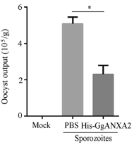
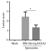
For the in vivo test, 1 × 10^6 sporozoites pretreated with rHis-GgANXA2 or PBS were administered to chickens via cloacal inoculation. At 5 days post-infection (dpi), cecal lesions and parasite burden were assessed, while oocyst output was monitored from 7 to 10 dpi, following established protocols [11, 25, 26].
RNA extraction, reverse transcription, and qPCR
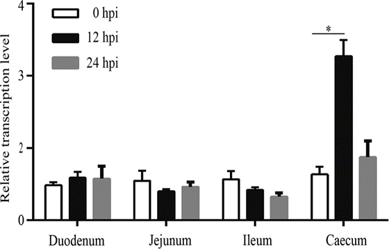
Chickens were orally challenged with 2 × 10^4 sporulated E. tenella oocysts per bird, and tissue samples from the duodenum, jejunum, ileum, and cecum were collected at 0, 12, and 24 hours post-infection. Total RNA was isolated using a Promega RNA purification kit (Madison, WI, USA). 1 μg of RNA was reverse transcribed to cDNA with M-MLV reverse transcriptase in the presence of RNase inhibitor. Quantitative PCR was conducted in triplicate using RealStar Green Fast Mixture (A303; GenStar, Beijing, China) on an ABI Q5 instrument. Threshold cycle values were normalized against GgGAPDH, and primers for GgANXA2 and GgGAPDH are listed in Table 1.
Table 1.
|
Gene |
Sense Primer (5’-3’) |
Antisense Primer (5’-3’) |
|
GgANXA2 |
AGGGCCTGGGAACTGATGAA |
GCCAGGGCAACCATTAGCTT |
|
GgGAPDH |
CACCGCTATTCCTTATAAAGAAAGT |
AAACACTTTCTTTATAAGGAATAGC |
Evaluation of protective effect of rHis-EtSERPIN1
Seven-day-old chickens were randomly assigned into three groups (n = 35 per group). The immunization procedure followed a previous protocol [11]. The EtSERPIN1 group received 50 μg of rHis-EtSERPIN1 emulsified in Freund’s complete adjuvant (FCA; Sigma) via subcutaneous injection. Groups PBS-I and PBS-II received PBS-FCA. Seven days later, chickens were boosted with either rHis-EtSERPIN1 in Freund’s incomplete adjuvant (FIA; Sigma) or PBS-FIA. One week post-booster, all chickens except PBS-I were orally challenged with 1 × 10^4 sporulated E. tenella oocysts per chicken.
Protective efficacy was determined using the anticoccidial index (ACI), calculated based on body weight gain, survival rate, oocyst output, and cecal lesion score. Weight gain was measured from 0 to 10 dpi, and oocyst output was quantified at 7 and 10 dpi. Cecal lesions were assessed in 8 chickens per group at 5 dpi, and ACI was calculated as:
ACI = (the relative body weight gain + survival rate) − (oocyst count index + lesion score index) [27].
Measurement of serum IgG and cecal sIgA
To evaluate immune responses, serum IgG and secretory IgA (sIgA) levels were quantified in each chicken group (n = 3) using an ELISA method, following established procedures [28]. Chickens were sacrificed by cervical dislocation, after which the ceca were carefully excised and cut along the longitudinal axis. The tissue samples were placed on ice in 10 mL of PBS, supplemented with 0.05 trypsin inhibitory units/mL aprotinin, 5 mM EDTA, 2 mM PMSF, and 0.02% NaN₃, and incubated for 4 hours to extract sIgA.
For the ELISA, microplates were coated with recombinant His-tagged EtSERPIN1 to detect IgG in serum and sIgA in cecal preparations. HRP-conjugated rabbit anti-chicken IgG antibodies served as the secondary detection reagent. Each measurement was conducted in triplicate, and optical density (OD) was recorded at 450 nm using a Biotek microplate reader.
Statistical procedures
All experiments were independently repeated three times, with three technical replicates per assay. Statistical analyses were conducted using GraphPad Prism 9.0. Comparisons between groups employed Student’s t-test, and results were reported as mean ± standard deviation (SD). A P-value < 0.05 was considered indicative of statistical significance.
Surface localization and functional role of EtSERPIN1
Previous studies reported the expression of EtSERPIN1 in sporozoites and schizonts [14], yet its presence during gametogony and exact localization on sporozoites were not fully defined. To clarify this, recombinant His-EtSERPIN1 with a molecular weight of approximately 50 kDa was produced (Figure 1a) (lane 1) and used to raise mouse polyclonal antibodies (pAb). Western blotting of sporozoite lysates with the anti-EtSERPIN1 pAb revealed two protein bands around 42 kDa and 45 kDa (Figure 1a) (lane 2).
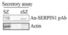
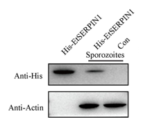
Analysis of secreted sporozoite proteins showed a band at ~42 kDa (Figure 1b), suggesting that EtSERPIN1 may exist in two molecular forms, with the lower band representing a secreted version lacking its signal peptide and cytoplasmic region. Using immunofluorescence assays, EtSERPIN1 was detected on the surface of sporozoites, as well as in schizonts and gametocytes (Figure 1c). These results indicate that EtSERPIN1 is both a membrane-bound and secreted protein, expressed across all life stages of E. tenella.
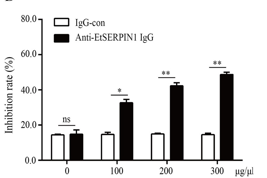
The ability of anti-EtSERPIN1 pAb to inhibit sporozoite invasion into host cells was confirmed in a dose-dependent assay. Pretreatment with 300 μg/mL anti-EtSERPIN1 blocked 48.77% of sporozoites from entering DF-1 cells, significantly higher than the 14.6% observed in controls (P < 0.05) (Figure 1d).
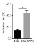
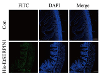
To further assess the role of surface EtSERPIN1, its adhesion and invasion properties were tested using a yeast surface display system and immunohistochemical assays. Yeast expressing surface EtSERPIN1 displayed enhanced adherence to DF-1 cells compared with control yeast (Figure 1f) (P < 0.05). Immunohistochemistry confirmed specific binding of recombinant His-EtSERPIN1 to cecal tissue sections, further supporting its adhesive function (Figure 1g).
|
|
|
|
|
a) |
||
|
|
||
|
b) |
c) |
d) |
|
|
|
|
|
e) |
f) |
g) |
|
Figure 1. |
||
Identification of GgANXA2 as an EtSERPIN1 binding partner
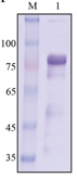
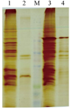
To explore host proteins interacting with EtSERPIN1, membrane proteins from chicken cecal epithelial cells were isolated and tested by GST pull-down combined with mass spectrometry (MS). Recombinant GST-tagged EtSERPIN1 was successfully expressed and purified (Figure 2a). SDS-PAGE analysis revealed several differential bands in the rGST-EtSERPIN1 pull-down compared to GST controls (Figure 2b).
Mass spectrometry identified 27 potential host proteins associated with EtSERPIN1. Subcellular localization predictions via WoLF PSORT and UniProt suggested that four proteins were membrane-localized, namely GgANXA2, GgANXA3, GgANXA5, and GgANXA13. Because parasite adhesion and invasion are mediated by interactions with host surface receptors, these four proteins were prioritized for further study.
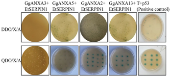
A point-to-point yeast two-hybrid (Y2H) assay was performed to verify interactions. Yeast co-transformed with pGBKT7-EtSERPIN1 and either pGADT7-GgANXA2 or pGADT7-GgANXA13 produced blue colonies on DDO/X/A and QDO/X/A plates, whereas yeast containing GgANXA3 or GgANXA5 constructs showed no color change (Figure 2c). This suggested that EtSERPIN1 may specifically interact with GgANXA2 and GgANXA13.
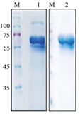
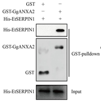
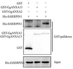
To confirm direct binding, a GST pull-down assay was conducted. Recombinant rGST-GgANXA2 and rGST-GgANXA13 proteins were expressed and purified (Figure 2d). The results showed that His-EtSERPIN1 was pulled down by rGST-GgANXA2, but not by the GST-only control (Figure 2e). No interaction was observed with rGST-GgANXA13 (Figure 2f). These findings demonstrate a specific interaction between EtSERPIN1 and GgANXA2, a chicken cecal membrane protein, implicating this interaction in parasite adhesion and invasion.
|
|
|
|
|
a) |
b) |
c) |
|
|
|
|
|
d) |
e) |
f) |
|
Figure 2. EtSERPIN1 Interaction with GgANXA2. a) SDS-PAGE analysis of the purified recombinant GST-tagged EtSERPIN1 (rGST-EtSERPIN1). b) SDS-PAGE of cecal epithelial membrane proteins bound to rGST-EtSERPIN1. M indicates protein markers. Lane 1: GST control expressed in E. coli BL21; Lane 2: pull-down of GST alone; Lane 3: rGST-EtSERPIN1 expressed in E. coli BL21; Lane 4: pull-down products of rGST-EtSERPIN1. c) Yeast two-hybrid assay to examine EtSERPIN1 binding with host proteins; p53 served as the positive control. d) SDS-PAGE of purified recombinant GST-GgANXA2 and GST-GgANXA13. e, f) GST pull-down experiments assessing the interaction between rHis-EtSERPIN1 and rGST-GgANXA2 (e) or rGST-GgANXA13 (f). Western blot using anti-His mAb and anti-GST mAb confirmed binding. |
||
Recombinant GgANXA2 associates with sporozoites
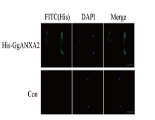
To verify EtSERPIN1’s binding partner, sporozoites were incubated with rHis-GgANXA2 and analyzed. Western blot results showed clear detection of rHis-GgANXA2 in sporozoites treated with the recombinant protein, whereas untreated sporozoites showed no detectable signal (Figure 3a). Immunofluorescence assays (IFA) using anti-His mAb demonstrated that rHis-GgANXA2 localized on the sporozoite surface (Figure 3b). These findings indicate a direct interaction between GgANXA2 and sporozoite surface proteins, confirming the association with EtSERPIN1.
|
|
|
|
a) |
b) |
|
Figure 3. Binding of rGgANXA2 to E. tenella sporozoites. Sporozoites were incubated with rHis-GgANXA2 and analyzed by western blot (A) and IFA (B) using anti-His mAb. Scale bars: 50 μm. |
|
Recombinant GgANXA2 and Anti-GgANXA2 block EtSERPIN1 adhesion
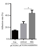
The functional role of GgANXA2 in mediating EtSERPIN1 adhesion was tested using a yeast surface display system. Yeast cells displaying EtSERPIN1 showed a reduced adhesion rate when pretreated with rGgANXA2 (44.32%) compared with the PBS control (26.75%) (P < 0.05) (Figure 4a).
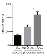
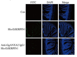
Pre-blocking DF-1 cells with anti-GgANXA2 antibody caused a further reduction in adhesion relative to cells incubated with IgG alone (Figure 4b). Immunohistochemical staining indicated that rHis-EtSERPIN1 binds specifically to cecal tissue. However, prior incubation with anti-GgANXA2 IgG markedly reduced this binding, suggesting that GgANXA2 is required for efficient EtSERPIN1-mediated adhesion (Figure 4c).
|
|
|
|
|
a) |
b) |
c) |
|
Figure 4. GgANXA2 and its specific antibody reduce EtSERPIN1 adhesion. a) Adhesion of yeast displaying EtSERPIN1 pretreated with rGgANXA2 or PBS. b) DF-1 cells pre-blocked with anti-GgANXA2 pAb or IgG were incubated with yeast expressing EtSERPIN1 or empty vector, and adhesion rates calculated. *P < 0.05. c) Cecal tissue pre-treated with anti-GgANXA2 pAb was incubated with rHis-EtSERPIN1, detected with anti-His mAb and FITC-conjugated secondary antibody. Nuclei stained with DAPI. Scale bars: 50 μm. |
||
Recombinant GgANXA2 limits sporozoite invasion in vitro and in vivo
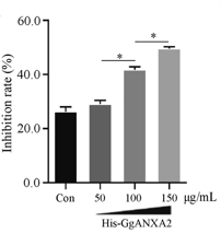
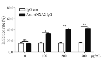
The effects of rGgANXA2 and anti-GgANXA2 antibody on sporozoite invasion were assessed in cell culture and live chickens. Pre-incubation with rGgANXA2 or blocking host cells with anti-GgANXA2 reduced sporozoite invasion in a dose-dependent manner (Figures 5a and 2b), confirming that both the recombinant protein and antibody interfere with parasite entry.
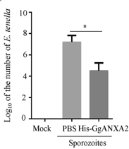
In vivo, chickens inoculated with sporozoites pretreated with rGgANXA2 showed lower cecal parasite burden (Figure 5c), fewer oocysts in feces (Figure 5d), and reduced cecal lesion scores at 5 dpi compared to controls (Figure 5e). These results demonstrate that GgANXA2 can effectively inhibit E. tenella sporozoite infection in both experimental settings.
|
|
|
|
|
a) |
b) |
|
|
|
|
|
|
c) |
d) |
e) |
|
Figure 5. rGgANXA2 and anti-GgANXA2 antibody inhibit sporozoite invasion. a) In vitro invasion of sporozoites treated with rGgANXA2 at different concentrations. b) DF-1 cells pre-incubated with anti-GgANXA2 pAb at varying doses were infected with sporozoites; invasion rates measured. c–e) In vivo infection: chickens received sporozoites pretreated with rGgANXA2. C. Cecal parasite load at 5 dpi. D. Fecal oocyst counts at 7–10 dpi. E. Cecal lesion scores at 5 dpi. *P < 0.05, **P < 0.01. |
||
Modulation of GgANXA2 transcription in the cecum during E. tenella infection
We investigated how GgANXA2 mRNA levels vary in different intestinal regions (duodenum, jejunum, ileum, and cecum) following E. tenella infection. qPCR results showed that at 12 h post-infection (hpi), transcription in the duodenum, jejunum, and ileum remained largely unchanged compared with baseline. In contrast, cecal GgANXA2 transcripts were significantly higher than at 0 hpi (Figure 6), suggesting that GgANXA2 upregulation in the cecum may be associated with sporozoite invasion.
|
|
|
Figure 6. Relative expression of GgANXA2 following E. tenella infection. qPCR quantified mRNA levels in duodenum, jejunum, ileum, and cecum at 0, 12, and 24 hpi. *P < 0.05 denotes statistically significant differences. |
Anti-E. tenella responses elicited by rHis-EtSERPIN1 immunization
Because EtSERPIN1 participates in parasite adhesion and invasion, we hypothesized that antibodies raised against rHis-EtSERPIN1 would interfere with E. tenella infection. Chickens were immunized and monitored for survival, weight gain, cecal lesions, and oocyst shedding. All groups survived (100% survival). Chickens receiving rHis-EtSERPIN1 exhibited significantly increased relative body weight compared to controls (Table 2). Cecal lesion scores were reduced (P < 0.05), and oocyst excretion was markedly lower in the immunized group. The anticoccidial index (ACI) for rHis-EtSERPIN1 reached 170.14, indicating moderate protective efficacy (Table 2). These findings support the role of rHis-EtSERPIN1 as an immunogenic candidate.
Induction of systemic and mucosal antibodies by rHis-EtSERPIN1
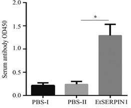
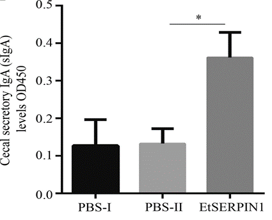
To evaluate humoral immune responses, we measured serum IgG and cecal sIgA specific to EtSERPIN1 7 days after the booster dose. Chickens immunized with rHis-EtSERPIN1 showed substantially higher serum IgG (Figure 7a) and elevated cecal sIgA (Figure 7b) compared with the PBS group, indicating that EtSERPIN1 stimulates both systemic and mucosal immunity.
Table 2. Protective efficacy of rHis-EtSERPIN1 against E. tenella. Values with different letters indicate significant differences (P < 0.05). ACI = (relative weight gain + survival rate) − (lesion score index + oocyst count index). Activity thresholds: excellent, ACI >180; moderate, 179>ACI>160; limited, 159>ACI>120; nonactive, ACI <120.
|
Treatment Group |
Survival Rate (%) |
Mean Weight Gain (g) |
Relative Weight Gain (%) |
Oocyst Output (× 10⁵/g) |
Oocyst Index |
Lesion Score |
Lesion Index |
Anticoccidial Index (ACI) |
|
PBS-I |
100 |
167.4 ± 4.85a |
100 |
0 |
0 |
0 |
0 |
200 |
|
PBS-II |
100 |
95.4 ± 4.33c |
56.99 |
6.83 ± 0.29b |
40 |
3.5 ± 0.16a |
35 |
81.99 |
|
EtSERPIN1 |
100 |
157.6 ± 4.95b |
94.14 |
3.03 ± 0.24a |
10 |
1.4 ± 0.1b |
14 |
170.14 |
|
|
|
|
a) |
b) |
|
Figure 7. EtSERPIN1-specific antibody responses. a) Serum IgG measured 7 days post-booster. b) Cecal sIgA determined at 10 dpi. *P < 0.05 indicates significant differences. |
|
Eimeria species in chickens progress through multiple developmental stages, with gene expression patterns typically reflecting the specific function required at each stage. Previous research has shown that EtSERPIN1 is expressed in oocysts, sporozoites, and merozoites [14], but its presence during gametogenesis was not previously determined. In this study, indirect immunofluorescence assays (IFA) demonstrated that EtSERPIN1 is also present during gamete development, indicating that it is expressed throughout the lifecycle of E. tenella and may possess multiple functional roles. Bioinformatics analyses identified both a signal peptide and a transmembrane domain, suggesting EtSERPIN1 functions as a membrane-associated protein [14]. Consistent with these predictions, we observed EtSERPIN1 localized on the sporozoite membrane, supporting its potential role in host cell adhesion and invasion.
The EtSERPIN1 coding sequence encodes a protein of approximately 45 kDa [14]. Interestingly, western blot analysis using anti-EtSERPIN1 antibodies revealed two bands with distinct molecular weights in sporozoite extracts, echoing previous findings for T. gondii SERPIN [19]. Evidence from T. gondii indicates that secreted SERPINs can protect host proteins from proteolytic degradation [19]. Accordingly, we hypothesize that immature EtSERPIN1 resides in the parasite cytoplasm as a larger, inactive form, while mature, cleaved EtSERPIN1 is secreted extracellularly to perform its function, appearing as a smaller band. Prior studies also detected EtSERPIN1 in host cells following sporozoite infection [15], and our findings indicate that the protein exhibits both surface-bound and secreted characteristics, suggesting its potential as a protease inhibitor.
The invasion of host cells by E. tenella, a highly pathogenic coccidian causing substantial economic losses in poultry, is a multifaceted process requiring several parasite proteins [29, 30]. Secretory proteins, particularly during early invasion stages, are critical in facilitating parasite entry. Prior work highlights the roles of rhoptry and microneme proteins in apicomplexan host invasion [30, 31]. Microneme proteins such as MIC2, MIC3, and MIC8 are central to sporozoite adhesion, while rhoptry neck proteins (RON2/4/5/8) interact with apical membrane antigen (AMA1) to mediate invasion [11, 12, 32–34]. Despite this knowledge, the invasion mechanisms in Eimeria spp. sporozoites remain poorly understood. Earlier studies reported elevated EtSERPIN1 expression in sporozoites relative to other stages [35] and localization at the apical end during invasion [15]. Consistent with these reports, our experiments demonstrated that anti-EtSERPIN1 antibodies reduced sporozoite invasion in a dose-dependent manner. Additionally, both immunohistochemistry and yeast surface display assays confirmed that EtSERPIN1 enhances parasite adhesion: yeast expressing EtSERPIN1 adhered more efficiently to host cells, and recombinant EtSERPIN1 bound cecal tissue, indicating a direct role in host cell attachment.
Protein-protein interactions are essential for the biological activity of invasion-related molecules [36]. Understanding these interactions can reveal the mechanisms of sporozoite invasion and identify novel antigen targets [37]. Prior studies on Eimeria invasion proteins, such as EtMIC8, EtCab, and EtCDPK4, underline their importance in sporozoite entry into host cells [11, 22, 38]. However, receptors on host epithelial cells facilitating adhesion and invasion are still poorly characterized. For instance, EaMIC3 from E. acervulina interacts with chicken UBE2F, promoting invasion [39], while EtMIC8-EGF binds GgE-PCAM on chicken epithelial cells, essential for attachment and entry [24]. Our findings extend this knowledge by confirming that EtSERPIN1 interacts with the host membrane protein GgANXA2, and that disruption of this interaction—via recombinant GgANXA2 or specific antibodies—significantly impairs sporozoite invasion, highlighting the EtSERPIN1-GgANXA2 complex as a key factor in host cell entry. These results suggest EtSERPIN1 is a promising therapeutic target for controlling coccidiosis, though further research is needed to map the precise binding domains responsible for GgANXA2 interaction.
Adhesion and invasion are critical for establishing infection by apicomplexan parasites. Host antibodies targeting these processes can inhibit sporozoite entry and reduce disease severity. For example, AMA1 and RON4 in Neospora canis generate potent antibodies against neosporosis, while Eimeria MIC2, MIC3, MIC8, and AMA1 elicit moderate-to-high anti-coccidial responses [11, 40–42]. In this study, immunization with recombinant EtSERPIN1 significantly enhanced body weight gain, reduced fecal oocyst shedding, and lowered cecal lesion scores. Correspondingly, serum IgG and cecal sIgA levels increased following immunization, indicating activation of systemic and mucosal immunity. sIgA, a frontline defense against intestinal parasites, has been shown to impair E. tenella development when present in cecal contents [43–45]. Therefore, elevated anti-EtSERPIN1 sIgA likely contributes to enhanced resistance against infection.
Conclusion
In conclusion, EtSERPIN1, a protease inhibitor from E. tenella, interacts with the host membrane protein GgANXA2 and is pivotal for sporozoite adhesion and invasion. Disruption of this interaction through recombinant GgANXA2 or antibodies inhibits parasite entry, while EtSERPIN1 immunization confers protective immunity. Targeting the EtSERPIN1-GgANXA2 axis may provide a novel strategy for coccidiosis control, warranting further studies to uncover its detailed molecular mechanisms and therapeutic applications.
Acknowledgments: None
Conflict of Interest: None
Financial Support: None
Ethics Statement: None